
Okayama University Medical Research Updates (OU-MRU) Vol.5
February 02, 2015
Source: Okayama University, Center for Public Information
For immediate release: 28 January 2015
Okayama University research: Cell injections get to the heart of congenital defects
(Okayama, 28 January 2015) Researchers at Okayama University and Okayama University Hospital show that children suffering from a condition known as hypoplastic left heart syndrome (HLHS) experienced some improvement in cardiac function in the months following injection of CDCs.
Children suffering from a lethal congenital heart defect may benefit from injections of a type of stem cells – cardiosphere-derived cells (CDCs). A study conducted by researchers at Okayama University and Okayama University Hospital showed that children suffering from a condition known as hypoplastic left heart syndrome (HLHS) experienced some improvement in cardiac function in the months following injection of CDCs.
The left ventricle, aorta and related valvular components in the hearts of HLHS patients are underdeveloped, with not enough cells being produced. Children born with the condition may die if they do not receive immediate medical attention. They require surgery within a few days of birth and additional treatment is needed long-term to deal with the right-ventricular-dependent circulation.
Previous studies had suggested that loss of a type of stem cell – progenitor cells which can differentiate into a range of other cells – is responsible for lower replication of the heart muscle cells. In their report of the research the Okayama researchers highlight that cardiac progenitor cells in children are “more abundant, self-renewing, and multipotent than those found in adults,” and propose their use as a safe therapeutic strategy for patients with heart failure.
The researchers monitored the heart functions in both a group of 7 patients that had received cell injection and a control group of 7 patients who had not. As they conclude, “Our prospective controlled study, the first paediatric phase 1 clinical trial of stem cell therapy for heart disease to our knowledge, suggests that intracoronary infusion of autologous cardiac progenitor cells is a feasible and safe approach to treat children with HLHS.”
Background
Cardiosphere-derived cells
Tissue-specific stem progenitor cells can be found in the adult mammalian heart. When cloned in suspension they form cardiospheres. The patient-derived CDCs used in the study were tested prior to use to confirm expression of cardiac transcription factors and normal chromosomes to avoid tumour formation after injection. Transcoronoray Injection was successfully achieved in all patients with no adverse effects.
Cardiac function monitoring
The heart functions monitored by the researchers included right ventricular ejection fraction (RVEF), end-systolic volume (volume of blood at the maximum contraction point, ESV), and end-diastolic volume (volume of blood when at the maximum filling point, EDV), stroke volume and cardiac output. In addition the levels a type of peptide - brain natriuretic peptide (BNP) were monitored. These peptides are secreted by the ventricles of the heart in response to excessive stretching of the heart muscle cells.
Many of the parameters measured showed improvements in the group that had received CDCs. In particular RVEF levels improved from 36.1 ± 7.5% at baseline to 42.7% ± 8.7% at 12 months. BNP levels were decreased 18 months after CDC injection compared with controls ((26.3 ± 28.5 pg/mL vs. 68.6 ± 42.4 pg/mL).
The researchers associated the decrease in heart failure status in CDC-treated patients with the decrease in BNP levels 18 months after CDC compared with the controls. In addition, right ventricular wall masses had decreased significantly and tissue growth was improved suggesting long term benefits 3-18m months after CDC injection.
Study conclusions and limitations
Due to the rarity of the disease and the difficulty in recruiting a sample population the study was small and non-randomized. With groups of just 7 patients the size of the study may limit the ability to draw definite conclusions as to the safety and effectiveness of the using CDC injections to treat HLHS patients. However the improved RVEP 18 months after CDC CDC injection provides a positive proof of concept for the treatment.
Further studies are required to endorse the results. In addition the cell biological mechanisms for enhancing cardiac function are still not understood and a prognostic cell-tracking system may be required as new translational medicine for children.
Acknowledgments
We are indebted to the patients, the parents of the patients who gave their consent to participate in this trial, and the staff of the cardiac care unit and the catheterization laboratory at Okayama University Hospital.
This study was supported by grants from the Ministry of Health, Labour and Welfare (to HO), the Ministry of Education, Culture, Sports, Science and Technology (to HO), and the Research Foundation of Okayama University Hospital (to SS and HO). The study funding source had no role in study design, data collection, data analysis and interpretation, or in the preparation of the manuscript.
Reference
Shuta Ishigami, Shinichi Ohtsuki, Suguru Tarui, Daiki Ousaka, Takahiro Eitoku, Maiko Kondo, Michihiro Okuyama, Junko Kobayashi, Kenji Baba, Sadahiko Arai, Takuya Kawabata, Ko
Yoshizumi, Atsushi Tateishi, Yosuke Kuroko, Tatsuo Iwasaki, Shuhei Sato, Shingo Kasahara,
Shunji Sano and Hidemasa Oh “Intracoronary autologous cardiac progenitor cell transfer in patients with hypoplastic left heart syndrome (TICAP): A prospective phase 1 controlled trial” ,Circulation Research, (2015)
DOI: 10.1161/CIRCRESAHA.116.304671
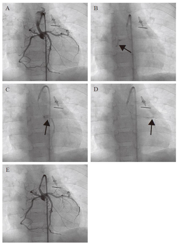
Figure 1. Angiographic images of transcoronary infusion of CDCs into children with HLHS. (A) Primary coronary angiography was performed prior to the CDC infusion. A 5 French guiding catheter Launcher (Medtronic) was placed in the hypoplastic ascending aorta, which was connected to the newly reconstructed aortic arch by the Norwood procedure. Selective CDC transfer was performed into right (B), left ascending (C), and left circumferential (D) coronary arteries by a stop-flow technique (arrows). (E) Hand injection of contrast medium demonstrates that the coronary arteries were unobstructed after CDC infusion.
Further information
Okayama University
1-1-1 Tsushima-naka , Kita-ku , Okayama 700-8530, Japan
Planning and Public Information Division, Okayama University
E-mail: www-adm@adm.okayama-u.ac.jp
Website: //www.okayama-u.ac.jp/index_e.html
Okayama Univ. e-Bulletin://www.okayama-u.ac.jp/user/kouhou/ebulletin/
Okayama University Medical Research Updates (OU-MRU)
Vol.1:Innovative non-invasive ‘liquid biopsy’ method to capture circulating tumor cells from blood samples for genetic testingVol.2:Ensuring a cool recovery from cardiac arrestVol.3:Organ regeneration research leaps forwardVol.4:Cardiac mechanosensitive integrator
About Okayama University
Okayama University is one of the largestcomprehensive universities in Japan with roots going back to the Medical Training Place sponsored by the Lord of Okayama and established in 1870. Now with 1,300 faculty and 14,000 students, the University offers courses in specialties ranging from medicine and pharmacy to humanities and physical sciences. Okayama University is located in the heart of Japan approximately 3 hours west of Tokyo by Shinkansen.