|
About the Center / Research Projects
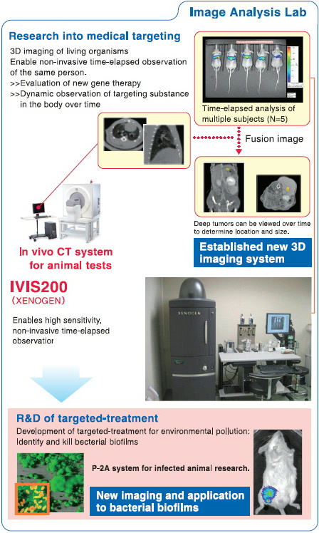
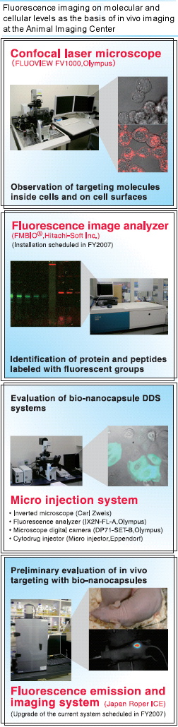
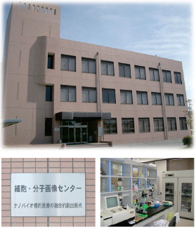
Our animal imaging center was
established in 2006 with the funding
from the Science and Technology
Promotion Bureau of MEXT.
Detection of metastatic cancer and
the monitoring of therapeutic drug
effects in animal models are the
primary projects currently ongoing
at this facility. Xenogen In-Vivo
Imaging System (IVIS), which detects
emissions from luciferase and other
fluorescent proteins, has been in
operation to analyze models for liver
cancer, malignant mesothelioma, and
prostatic cancer. A CT scanner, which
is expected to offer more accurate
3-D information, will be introduced
some time this year.
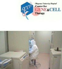
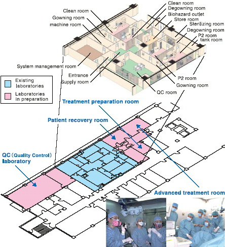
Role within ICONT
The animal imaging center is the main
facility of ICONT’s nanobio project.
The highly sensitive and accurate in
vivo imaging equipment is essential
for the development of successful
targeted therapy. Indispensable for all
of the nanobio-related projects
(cancer gene therapy using viral
vectors and non-viral vectors including
polymer, researches for bacterial biofilm,
etc.) at ICONT, the facility is expected
to become one of the main imaging
facilities for exploratory medical research
in Japan.
Address 2-5-1, Shikata-cho, Okayama 700-8558, Japan
|