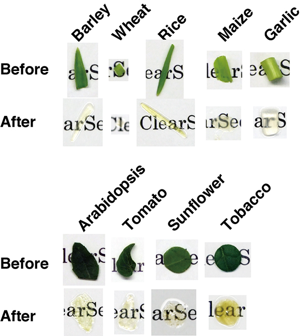
Leaves of five monocot and four dicot species were cleared by the ePro-ClearSee method. Leaves before and after clearing are indicated.
Enlarge Image
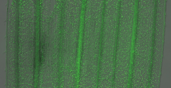
Signals of methylated histone (green) were shown on a bright field image of a wheat leaf.
Enlarge Image
Signals of methylated histone (green) and centromere specific histone variant (red) were shown on DAPI stained nuclei (gray).
Enlarge Image
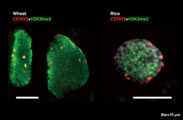
Signals of methylated histone (green) and centromere specific histone variant (red) were shown on DAPI stained nuclei (gray).
Enlarge Image
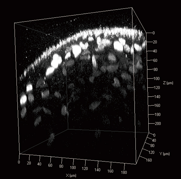
Signals of methylated histone (gray) were shown.
Enlarge Image
Quick immunohistochemistry with clearing detects epigenetic changes
Epigenetic changes in plant tissues are detected using a comparatively fast immunohistochemical method developed at Okayama University in Japan
Some inheritable traits stem from changes in how genes are expressed without affecting the DNA sequence, such as modifications to histones, the proteins responsible for ordering DNA into structural units or ‘nucleosomes’ that fit into cells. Okayama University researchers have now developed a technique that is sensitive enough to detect epigenetic changes while avoiding some of the drawbacks of previous techniques, such as poor sample penetration and lengthy preparation times.
The researchers based their approach on immunohistochemical staining, which uses the selective binding of antigens to specific proteins for imaging. Previous attempts with immunohistochemical staining have met with limited success due to autofluorescence interfering with signals and poor antibody permeability into the cells. One previous attempt successfully removed lipids, colours and cell walls, allowing antibodies access to the cells without errant autofluroescence, but took 7-9 weeks to complete and only proved successful in detecting the most abundant cell protein, which has a far stronger immunosignal than the targeted epigenetic modifications.
Kiyotaka Nagaki, Naoki Yamaji and Minoru Murata combined the use of enzymes (e) to digest cell walls and aid permeability with 2-propanol (Pro) and a “ClearSee” clearing treatments to make the leaf samples transparent (ePro-ClearSee). They demonstrated the technique on a range of samples including dicot and monocot species as either whole leaves, perforated leaves, cropped disc shapes and strips, identifying pros and cons for the different leaf shapes for different studies.
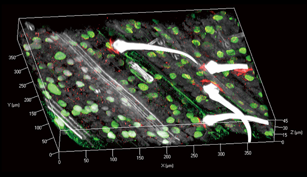
“ePro-ClearSee method enabled the detection of immunosignals 200 μ m deep in plant tissues without the need for sectioning for a short period of time,” they explain in their report of the technique, concluding that it may be applicable to other types of analysis as well.
Publication and Affiliation
Kiyotaka Nagaki*, Naoki Yamaji and Minoru Murata Investigations into ePro-ClearSee: a simple immunohistochemical method that does not require sectioning of plant samples 2017, Scientific Reports 7 42203
Institute of Plant Science and Resources, Okayama University, Kurashiki 710-0046, Japan.
*Correspondence: nagaki@rib.okayama-u.ac.jp